MyLab™Twice
1401-04-11
MyLab™X7
1401-04-12
MyLab™Class C

MyLab™Class C: Discover The Power Of Touch
Your Comfort With A Touch
Whenever physicians think of a high-level cardiovascular and general imaging ultrasound systems, they ask for up-to-date platforms, with high-performance and advanced on-board technologies as well as simplicity and ease of use. MyLab™Class C has been designed based on these key concepts to deliver a reliable diagnosis, and ensure every day productivity. With just one glance, you will understand how MyLab™Class C‘s simplicity has never been seen on such a high-level ultrasound scanner.
MyLab™Class C: Performance, Simplicity And Ergonomics With A Single Touch
High performance does not always mean large and stationary systems. A particular effort has been made to reduce size and increase the new MyLab™Class C’s ergonomics. This has led to a compact and agile system, which is easy to move and is able to adapt to any kind of environment, including most critical ones such as interventional and operating rooms. The height-adjustable and rotating keyboard, as well as the multiplane-articulated monitor arm, allow for optimal positioning at all times.

Opti-Light: High-Performance Optimized Lighting
Optimal lighting has always been a crucial factor for ultrasound imaging. The latest LCD Monitor Technology allows images to be clearly displayed under any condition. MyLab™Class C also introduces an additional unique feature: Opti-Light. This feature, thanks to a light point behind the monitor, allows the operator to control the room's lighting level directly from the system, through the especially designed controls located on the touch screen. Optimized working conditions increase users’ comfort and improve patient care.


High-Quality Touch Screen: Completeness With A Touch
The large high-quality touch screen is well positioned near the most important working area of the control panel. This touch-screen allows all mode-dependent parameters to be clearly displayed and changed with one simple touch.
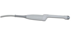
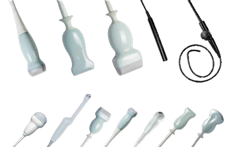
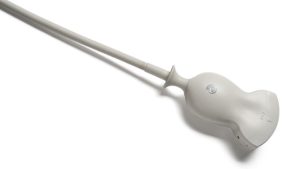
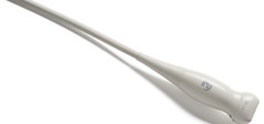
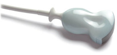
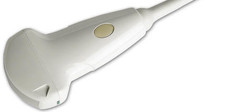
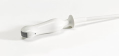
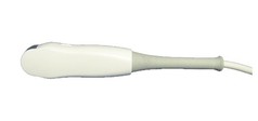
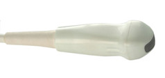
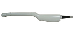
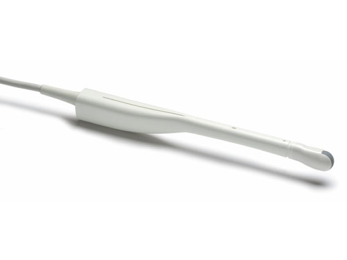
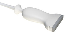
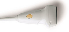
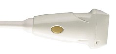
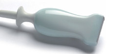
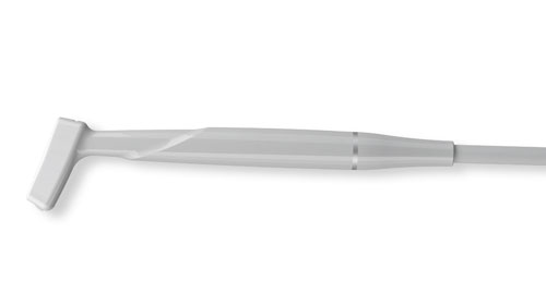
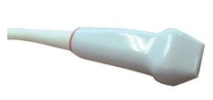
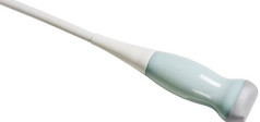
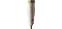
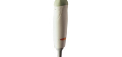
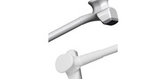
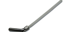
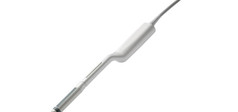
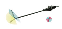
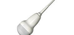
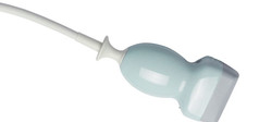
iQProbes: A Wide Range Of High-Tech Transducers
The primary component in the Signal Processing Chain leading to the final ultrasound diagnostic image is the transducer. The material design and technology employed to manufacture an ultrasound transducer are the key factors in determining the system’s image quality. iQProbes represent Esaote’s state-of-the-art Technology thanks to its innovative gold standard ultrasound transducers. Designed to improve performance and ergonomics, iQProbe Technology is based upon:
- Innovative Active Matrix Composite Material
- Multiple Adaptive Layers Solution
- Structure Filling Material Manufacturing Process
- Intelligent Geometric Lens Manufacturing Process

Integration With A Touch
Data management is very important today, both for users' comfort and patient care. Esaote offers an efficient solution for any need and any environment, ranging from stand-alone workstations up to complex modular architectures. MyLab™Desk represents a flexible way to connect your MyLab™ to the PC, easily! MyLab™Desk is Esaote’s answer to its user’s need for a simple and straightforward way to archive, review, post-process, report or print their MyLab™ examinations on a PC from the comfort of their (home) office or while travelling. MyLab™Desk provides the means to increase workflow and productivity in private offices, as well as in clinics and hospital departments.
- Archive, Review And Post-Process Examinations Performed With The MyLab™ Ultrasound Systems
- Import Esaote Native File Formats (UAF And EAF Raw Data) Via USB, CD/DVD And Network
- Perform General And Application-Specific Measurements
- Review, Change And Print The Examinations (Reports And Images)
- Export Data Using PC’s Standard Features, I.E. Burn On A CD/DVD, Email, Etc
Advanced Technologies For Every Clinical Need
The unique “Twice Ultrasound Vision” with eHD Technology offers the latest innovations for diagnostic confidence and optimized workflow, combining top performance and exclusive solutions to change daily clinical outcomes.
 Click the image to view Esaote Advanced Technologies Images
Imaging Processing
Esaote offers many technologies for imaging enhancement. With TEI™, the harmonic signal is fully preserved without degradation of the acoustic information. MView and XView improve the quality of ultrasound images by reducing the presence of artifacts, shadowing and speckle.
Raw data post-processing: allows post-processing of images and video clips previously acquired and saved into the archive. This feature is very helpful in the clinical workflow and provides physicians with image optimization in the off-line stage.
XFlow – Extraordinary flow sensitivity and spatial resolution: XFlow provides direct visualization of blood echoes, extending wideband resolution, high frame rates and wide dynamic range of blood flow.
CnTI™ – Contrast Tuned Imaging: Esaote’s proprietary CnTI™ (Contrast Tuned Imaging) provides high performance contrast enhanced ultrasound imaging with secondgeneration contrast media.
ElaXto – Further step towards tissue characterization: Non-invasive method to support the physician in assessing tissue elasticity.
 Click the image to view Esaote Advanced Technologies Images
X4D X3D: Esaote’s volumetric technology takes full advantage of the touch panel to optimize workflow and ease of use, and represents a breakthrough in technology.
XLight – A New Era in Volume Imaging: The new XLight technology guarantees to immediately reach amazingly realistic volumetric images also with very difficult to scan patients, both for gynecology as well as obstetrics exams. Lack of amniotic fluid, baby stage or position are no longer a barrier for providing a wonderful memory of the pregnancy. XLight provides the maximum volume rendering for all the clinical needs.
>> DOWNLOAD PDF BROCHURE [275 KB]
Virtual Biopsy – Advanced Biopsy also in very difficult approaches: Virtual Biopsy allows physicians to follow percutaneous procedures by superimposing needle tracking information on the real-time ultrasound image.
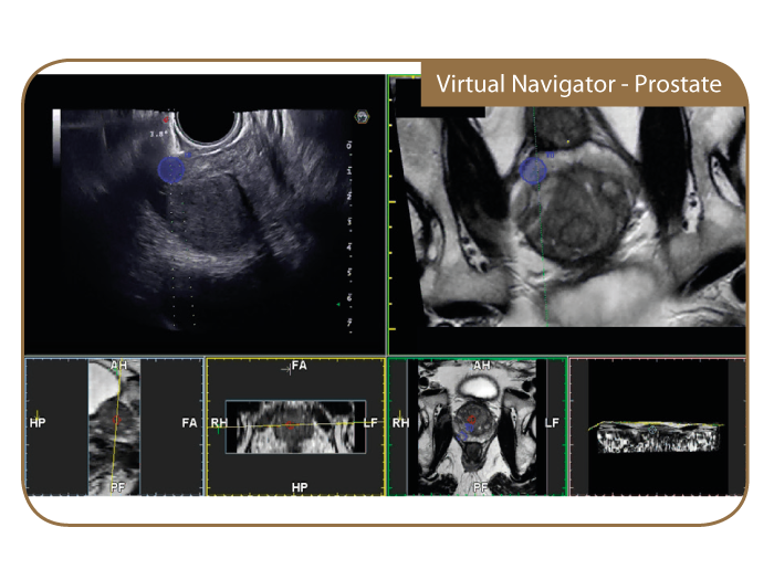
Virtual Navigator advanced tool for Fusion Imaging (US & CT/MR/PET-CT): Virtual Navigator, with fusion imaging applied to US enhances the information produced by an Ultrasound Scanner by combining images with a second modality (CT, MR, PET or 3D US) in real-time: yielding all the benefits of different modalities in the same exam.
>> GET ACCESS AND ENJOY VIRTUAL NAVIGATOR APP
XStrain4D: XStrain™ is a non-invasive tool to better investigate myocardial function and explore and quantify aspects of the heart’s physiology which were not possible to detect and quantify with previous ultrasound technologies.
>> GET ACCESS AND ENJOY XStrain™ APP
RFQIMT – RFQAS – Innovation and Accuracy in Vascular Imaging: The measurements, based on beyond state of the art RF-data technology, are real-time, accurate and provide measurement quality indicators overlaid on the B-mode ultrasound image.
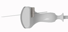
C 1-8

L 4-15 appleprobe
PA250
CA541 appleprobe
CA631
SC3123
SC3121
CA123
EC1123
EC123
LA533 appleprobe
LA523
LA435

SL2325
LA332 appleprobe
IH 6-18
SL3116
PA240
PA122
PA023
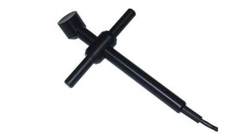
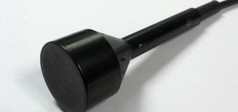
Pencil CW 2
Pencil HF CW
Pencil CW 5
S2MPW
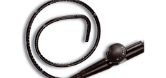
TEE022 Multiplane
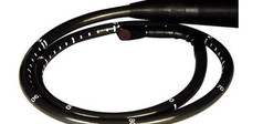
TEE132 Multiplane
SI2C41 appleprobe
IOT342 appleprobe
IOE323
TRT33 Bi-plane
LP323
BC441
BL433 appleprobe